Dental Implants (દાંતનું પ્રત્યારોપણ)
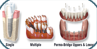
A dental implant is a "root" device, usually made of titanium, used in dentistry to support restorations that resemble a tooth or group of teeth to replace missing teeth.
Virtually all dental implants placed today are root-form endosseous implants, i.e., they appear similar to an actual tooth root (and thus possess a "root-form") and are placed within the bone (endo- being the Greek prefix for "in" and osseous referring to "bone"). The bone of the jaw accepts and osseointegrates with the titanium post. Osseointegration refers to the fusion of the implant surface with the surrounding bone. Dental implants will fuse with bone; however, they lack the periodontal ligament, so they will feel slightly different from natural teeth during chewing.
Prior to the advent of root-form endosseous implants, most implants were either blade endosseous implants, in that the shape of the metal piece placed within the bone resembled a flat blade, or subperiosteal implants, in which a framework was constructed to lie upon and was attached with screws to the exposed bone of the jaws.
What Are Dental Implants?
Dental implants are replacement tooth roots. Implants provide a strong foundation for fixed (permanent) or removable replacement teeth that are made to match your natural teeth.
What Are the Advantages of Dental Implants?
Improved appearance.
Dental implants look and feel like your own teeth. And because they are designed to fuse with bone, they become permanent.
Improved speech.
With poor-fitting dentures, the teeth can slip within the mouth causing you to mumble or slur your words. Dental implants allow you to speak without the worry that teeth might slip.
Improved comfort.
Because they become part of you, implants eliminate the discomfort of removable dentures.
Easier eating.
Sliding dentures can make chewing difficult. Dental implants function like your own teeth, allowing you to eat your favorite foods with confidence and without pain.
Improved self-esteem.
Dental implants can give you back your smile and help you feel better about yourself.
Improved oral health.Dental implants don't require reducing other teeth, as a tooth-supported bridge does. Because nearby teeth are not altered to support the implant, more of your own teeth are left intact, improving long-term oral health. Individual implants also allow easier access between teeth, improving oral hygiene.
Durability. Implants are very durable and will last many years. With good care, many implants last a lifetime.
Convenience. Removable dentures are just that; removable. Dental implants eliminate the embarrassing inconvenience of removing dentures, as well as the need for messy adhesives to keep them in place.
How Succssesful Dental Implants
Success rates of dental implants vary, depending on where in the jaw the implants are placed but, in general, dental implants have a success rate of up to 98%.
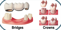
Ceremic Crown and Bridges (સિરામિક દાંતના કવર)
Crown and bridge treatment is a long-term method of replacing missing teeth.
A crown is placed on an individual tooth, where there is no sufficient tooth structure remaining to place a filling.
A bridge is applied when in space where one or more teeth have been lost. The teeth on either ends are crowned, and are referred to as abutments. The false teeth in a bridge that join the abutments are termed as pontics. Crowns and bridges are mostly manufactured from superior materials such as gold, porcelain, or a combination of metal fused to porcelain.
Both appearance and the function are considered when selecting the material that is best for you.
Wht is Crown?
A crown, often called as a cap, is an artificial hollow cover resembling a natural tooth crown that restores a decayed or damaged tooth to its normal shape and size. It is fabricated out of metal or ceramic material by special procedure in a dental lab.
Where can Crowns br Placed?
All the teeth that have had root canal treatments done should be covered with crowns to prevent their breakage as they have lost their natural hydrating mechanism and tend to be brittle.
Teeth that have been restored more than half by filling material should be crowned.
Tooth with bad aesthetics can be reshaped with crown to improve the aesthetics.
Damage to the tooth structure because of wear and tear can be restored with crown.
Fractured tooth can be restored with crown.
Teeth with wide spaces between them resulting in food lodgment or unpleasant appearance can be crowned. What
What is Bridge?
Dental bridge is a false tooth or teeth, known as a pontic, which is fused between two porcelain crowns to fill in the area left by a missing tooth or teeth. The two crowns holding it in place that are attached onto your teeth on each side of the false tooth. This is known as a fixed bridge. This used to replace one or more missing teeth. procedure is Fixed bridges cannot be taken out of your mouth as you might do with removable partial dentures. Bridges can reduce your risk of gum disease, help correct some bite issues and even improve your speech. Bridges require your commitment to serious oral hygiene, but will last as many ten years or more.
Dental Bridges are used to replace one or more missing teeth by taking the support of the adjacent natural teeth. Putting a dental bridge avoids problems like rotation or shifting of adjacent teeth which results in disturbed bite, gum diseases, decay of adjacent teeth and supra eruption of opposite tooth.
Disimpaction (ડહાપણની ડાઢની સર્જરી)
Bleaching & Composite (દાંત સફેદ બનાવવા)
Diamond (દાંતમાં હીરો લગાવવો)
Orthodontic Treatment (વાંકા ચુંકા દાંતની સારવાર)
Dental Beauty Treatment & Cosmetic Treatment (નાના બાળકોના દાંતની સારવાર)
Time Require: 2 seatings
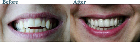
Cosmetic dentistry is generally used to refer to any dental work that improves the appearance (though not necessarily the function) of a person's teeth, gums and/or bite. Many dentists refer to themselves as "cosmetic dentists" regardless of their specific education, specialty, training, and experience in this field.
Cosmetic dentistry may involve:
The addition of a dental material to teeth or gums - examples: bonding, porcelain veneers (laminates), crowns (caps), gum grafts
The removal of tooth structure or gums - examples: enameloplasty, gingivectomy
Neither adding nor removing dental materials, tooth structure, or gums - examples: teeth whitening (bleaching), gum depigmentation
Straightening of teeth accompanied by improvement in appearance of face - Orthodontics
Whitening, or "tooth bleaching", is the most common cosmetic dental procedure. While many whitening options are now available, including over the counter products, dentist-supervised treatments remain the recommended procedures for lightening discolored teeth.
Tooth reshaping removes parts of the enamel to improve the appearance of the tooth. It may be used to correct a small chip, or to alter the length, shape or position of teeth; it can be used to correct crooked or excessively long teeth. The removed enamel is irreplaceable, and may sometimes expose dentin. It is also known as enameloplasty, odontoplasty, contouring, recontouring, slenderizing, stripping or sculpting. This procedure offers fast results and can even be a substitute for braces under certain circumstances.
Complete Denture Prosthesis (દાંતના તમામ પ્રકારના ચોકઠાં બનાવા)
It improves imperfection of bite and enhances the appearance of smile by correcting the entire dentition which is deteriorated due to several factors.
It is basically a combination of various dental procedures to primarily improve the functionality of teeth in terms of chewing, talking and biting.

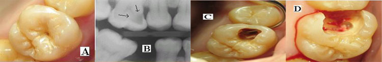
Filling of Decayed Teeth
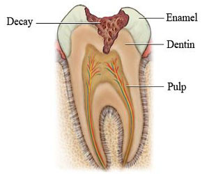
What is tooth decay?
Tooth decay is the process that results in a cavity (dental caries). It occurs when bacteria in your mouth make acids that eat away at a tooth. If not treated, tooth decay can cause pain, infection, and tooth loss.
Tooth decay is damage to the enamel of your teeth. It leads to cavities that can affect every layer of the tooth. Tooth decay occurs when acids produced by bacteria in the mouth eat away at a tooth. Any part of a tooth can decay, from the roots below the gum line to the chewing surface.
You can easily prevent tooth decay by brushing and flossing your teeth regularly, seeing your dentist for teeth cleaning and checkups, and avoiding foods that are high in sugar.
How is it treated?
Treatment for tooth decay depends on how bad it is. You may be able to reverse slight tooth decay by using fluoride. To fix cavities caused by mild tooth decay, your dentist will fill the cavities with another substance (fillings). For more severe tooth decay, you may need a crown or root canal. In extreme cases, your dentist may have to remove the tooth.
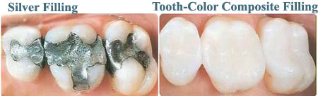
Types of fillings:
(1) Silver fillings
(2) Tooth-Color composition fillings
Flap Surgery (પેઢાના રોગોની સારવાર)
By far the most common surgery used in periodontal therapy is flap surgery. Moderate to advanced periodontal disease involved gum pockets (See What Is Periodontal Disease?) that are too deep to clean without reflecting back the gum tissue for access. Without this access, deep calculus and plaque cannot be removed from the root, and the disease will progress. This surgical cleaning procedure is often called open flap curettage.
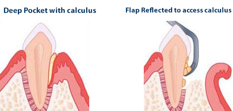
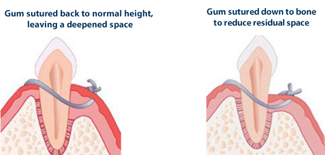
Once the pocket is cleaned, the gum may be returned to its original level. This results in a clean root, but the deepened space is still present. Frequent cleanings by the hygienist are necessary to remove the plaque in the residual pocket that the patient cannot reach with flossing and brushing
Even when there is good oral hygiene and regular quarterly recalls, the bacteria may still continue to cause the pocket to become reinfected. When cosmetics are not a concern (on the lower teeth, the inside of the upper teeth, and the outside of the upper back teeth), the surgeon may elect to suture the gum down to where the bone has resorbed, reducing the depth of the space. If the space is reduced to 3 millimeters or less, the patient is able to reach the bottom of the space with daily brushing and flossing, eliminating the disease.
In summary, for most moderate and advanced cases, it is important to be able to reach far under the gum to treat the infection and diseased tissues.
By using flap surgery, the periodontist is able to access these areas to provide the optimal care available. With today's medications, surgery should be painless with only a minimal amount of post-operative discomfort
Root canal treatment (દાંતના મૂળામાં સડો અને તેની સારવાર)
What is Root Canal?
A root canal is a treatment used to repair and save a tooth that is badly decayed or becomes infected. During a root canal procedure, the nerve and pulp are removed and the inside of the tooth is cleaned and sealed. Without treatment, the tissue surrounding the tooth will become infected and abscesses may form.
"Root canal" is the term used to describe the natural cavity within the center of the tooth. The pulp or pulp chamber is the soft area within the root canal. The tooth's nerve lies within the root canal.
A tooth's nerve is not vitally important to a tooth's health and function after the tooth has emerged through the gums. Its only function is sensory -- to provide the sensation of hot or cold. The presence or absence of a nerve will not affect the day-to-day functioning of the tooth.
Root canal treatment Steaps:
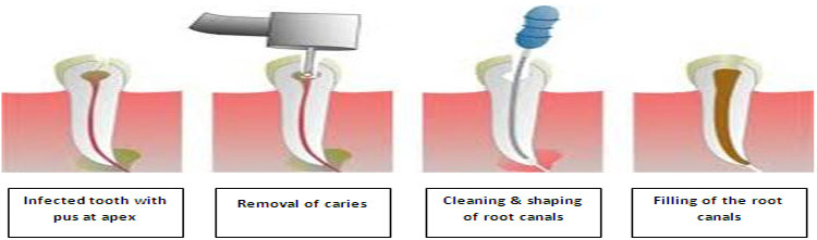
First, an opening is made through the back of a front tooth or the crown of a molar or pre-molar.
After the diseased pulp is removed (a pulpectomy), the pulp chamber and root canals are cleaned, enlarged and shaped in preparation for being filled.
If more than one visit is needed, a temporary filling is placed in the crown opening to protect the tooth between dental visits.
The temporary filling is removed and the pulp chamber and root canal permanently filled. A tapered, rubbery material called gutta-percha is inserted into each of the canals and is often sealed into place with cement. Sometimes a metal or plastic rod is placed in the canal for structural support.
In the final step, a crown is usually placed over the tooth to restore its natural shape and appearance. If the tooth is very broken down, a post may be required to build it up prior to placing a crown.
Intraoral Radiography (Intra Oral Camera અને Computer દ્વારા દાંતના તપાસ)
Dental Radiographs are commonly called x-rays. Dentists use radiographs for many reasons: to find hidden dental structures, malignant or benign masses, bone loss, and cavities.
A radiographic image is formed by a controlled burst of X-ray radiation which penetrates oral structures at different levels, depending on varying anatomical densities, before striking the film or sensor. Teeth appear lighter because less radiation penetrates them to reach the film. Dental caries, infections and other changes in the bone density, and the periodontal ligament, appear darker because X-rays readily penetrate these less dense structures. Dental restorations (fillings, crowns) may appear lighter or darker, depending on the density of the material.
The dosage of X-ray radiation received by a dental patient is typically small (around 0.150 mSv for a full mouth series, according to the American Dental Association website), equivalent to a few days' worth of background environmental radiation exposure, or similar to the dose received during a cross-country airplane flight (concentrated into one short burst aimed at a small area). Incidental exposure is further reduced by the use of a lead shield, lead apron, sometimes with a lead thyroid collar. Technician exposure is reduced by stepping out of the room, or behind adequate shielding material, when the X-ray source is activated.
Once photographic film has been exposed to X-ray radiation, it needs to be developed, traditionally using a process where the film is exposed to a series of chemicals in a dark room, as the films are sensitive to normal light. This can be a time-consuming process, and incorrect exposures or mistakes in the development process can necessitate retakes, exposing the patient to additional radiation. Digital x-rays, which replace the film with an electronic sensor, address some of these issues, and are becoming widely used in dentistry as the technology evolves. They may require less radiation and are processed much quicker than conventional radiographic films, often instantly viewable on a computer. However digital sensors are extremely costly and have historically had poor resolution, though this is much improved in modern sensors.
Placing the radiographic film or sensor inside the mouth produces an intraoral radiographic view.
Periapical view
The periapical view is taken of both anterior and posterior teeth. The objective of this type of view is to capture the tip of the root on the film. This is often helpful in determining the cause of pain in a specific tooth, because it allows a dentist to visualize the tooth as well as the surrounding bone in their entirety. This view is often used to determine the need for endodontic therapy as well as to visualize the successful progression of endodontic therapy once it is initiated. It can be used in case of detection hyperdontia(supernumerary teeth) & impacted teeth.
The name periapical is derived from the Greek peri, which means "around," and apical, which means "tip."
Bitewing view
The bitewing view is taken to visualize the crowns of the posterior teeth and the
height of the alveolar bone in relation to the cementoenamel junctions, which are
the demarcation lines on the teeth which separate tooth crown from tooth root. Routine
bitewing radiographs are commonly used to examine for interdental caries and recurrent
caries under existing restorations. When there is extensive bone loss, the films
may be situated with their longer dimension in the vertical axis so as to better
visualize their levels in relation to the teeth. Because bitewing views are taken
from a more or less perpendicular angle to the buccal surface of the teeth, they
more accurately exhibit the bone levels than do periapical views. Bitewings of the
anterior teeth are not routinely taken.
The name bitewing refers to a little
tab of paper or plastic situated in the center of the X-ray film, which when bitten
on, allows the film to hover so that it captures an even amount of maxillary and
mandibular information.
Occlusal view
The occlusal view is indicated when there is a desire to reveal the skeletal or
pathologic anatomy of either the floor of the mouth or the palate. The occlusal
film, which is about three to four times the size of the film used to take a periapical
or bitewing, is inserted into the mouth so as to entirely separate the maxillary
and mandibular teeth, and the film is exposed either from under the chin or angled
down from the top of the nose. Sometimes, it is placed in the inside of the cheek
to confirm the presence of a sialolith in Stenson's duct, which carries saliva from
the parotid gland.
The occlusal view is not included in the standard full mouth
series.
Full mouth series
A full mouth series is a complete set of intraoral X-rays taken of a patients' teeth
and adjacent hard tissue.
The Faculty of General Dental Practice of the Royal
College of Surgeons of England publication Selection Criteria in Dental Radiography
holds that given current evidence full mouth series are to be discouraged due to
the large numbers of radiographs involved, many of which will not be necessary for
the patient's treatment. An alternative approach using bitewing screening with selected
periapical views is suggested as a method of minimising radiation dose to the patient
while maximizing diagnostic yield.
Tooth Jewellery
What is tooth jewellery
Tooth Jewellery is the latest fashion craze to hit the UK. Tooth jewellery classic designs are an elegant compliment to standard jewellery. When placed on the tooth, the jewellery creates a distinctive expression of one’s individuality.
There are 2 main types of jewellery available
Twinkles
This is a collection of 24-carat gold and white gold jewellery. The jewellery is available in over 50 different designs with some including diamonds, sapphires and rubies.
Dental Gems
Are a range of glass crystals are available in nine different colours – diamond, rainbow, ruby, sapphire, emerald, emerald green, aquamarine, pink, sapphire light.
is the jewellery for males or females?
Both – there are many different designs which appeal to everyone. Children under the age of 16 should have parental consent.